How does diabetes damage the eyes?
High blood glucose levels damage the tiny capillaries that supply the retina - the light-sensitive tissue at the back of the eye. Over time, these vessels become leaky, blocked or grow abnormally. The result is diabetic retinopathy (DR), which can progress to severe vision loss or blindness if untreated.
India has over 77 million people with diabetes - one of the highest burdens in the world. Approximately 17 million have some form of diabetic retinopathy. Yet because the disease is painless in its early stages, many cases go undetected until vision is already compromised.
The stages of diabetic retinopathy
| Stage | What is happening | Symptoms | Treatment |
|---|---|---|---|
| Mild NPDR | Small balloon-like swellings (microaneurysms) in retinal vessels | None | Monitor every 12 months; optimise blood sugar |
| Moderate NPDR | More vessels blocked; dot and blot haemorrhages; hard exudates | Usually none; possible mild blurring | Monitor every 6 months; tighten glycaemic control |
| Severe NPDR | Many blocked vessels; venous beading; IRMA | May notice floaters or blurring | Consider panretinal laser; monitor every 3 months |
| PDR (Proliferative) | New fragile vessels growing on retina/disc; risk of haemorrhage | Sudden floaters, vision loss, dark curtain | Urgent panretinal photocoagulation ± anti-VEGF ± vitrectomy |
| DMO (any stage) | Fluid leak into macula causing swelling; distorts central vision | Blurred or distorted central vision | Anti-VEGF injections ± macular laser |
NPDR = Non-Proliferative Diabetic Retinopathy; PDR = Proliferative Diabetic Retinopathy; DMO = Diabetic Macular Oedema; IRMA = Intraretinal Microvascular Abnormalities.
Diabetic Macular Oedema (DMO): the most common cause of vision loss
The macula is the central part of the retina responsible for reading and fine detail. When diabetic vessels leak fluid into the macula, it swells - a condition called diabetic macular oedema. DMO can occur at any stage of diabetic retinopathy and is the single most frequent cause of diabetes-related vision impairment.
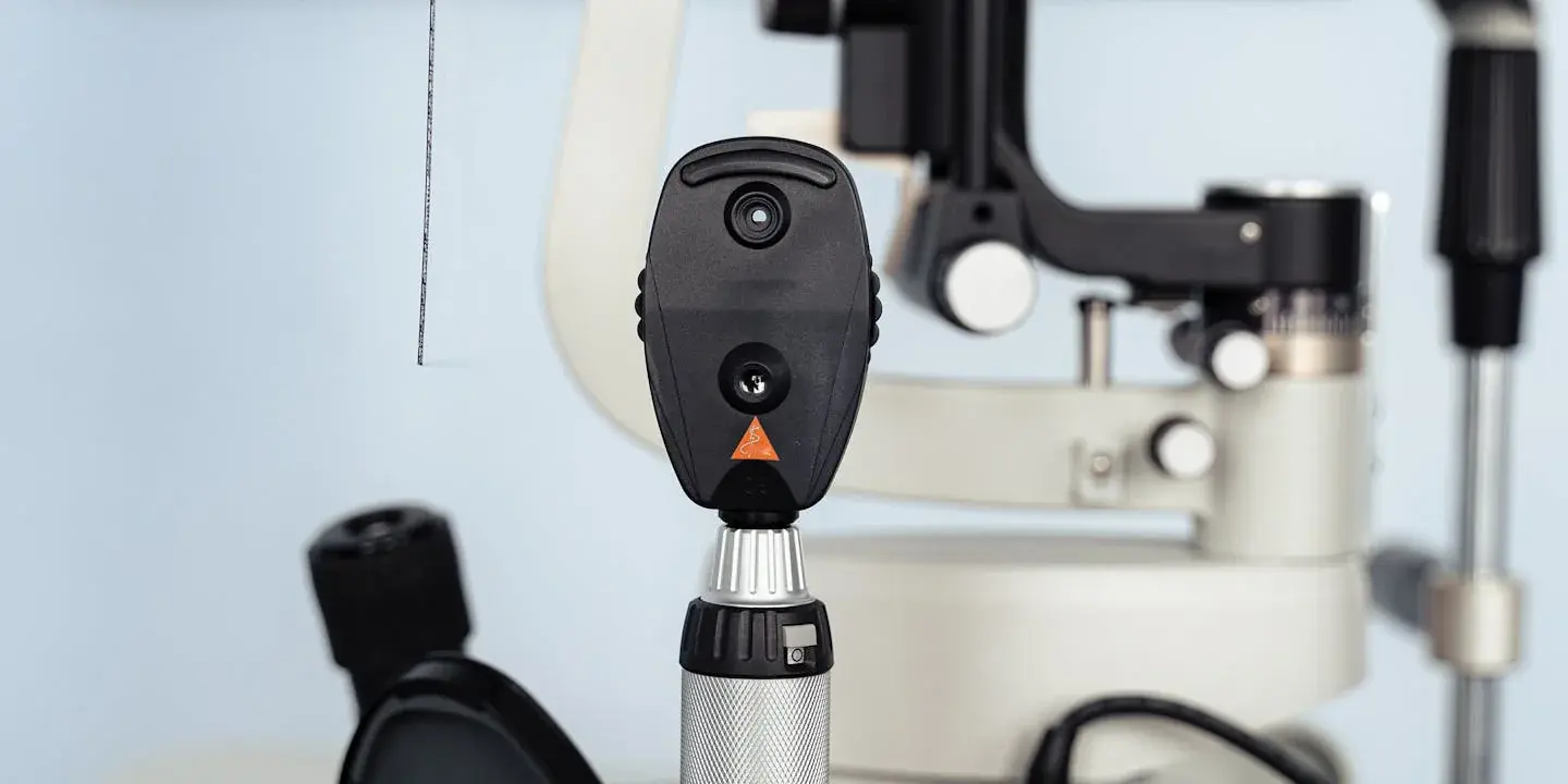
Modern OCT (optical coherence tomography) imaging allows precise measurement of macular thickness, enabling early detection and treatment monitoring. Eye Veda uses high-resolution OCT at every diabetic retina examination.
Treatment options for diabetic retinopathy
Anti-VEGF injections (ranibizumab, aflibercept, bevacizumab) are the gold standard for DMO. They block the protein (vascular endothelial growth factor) that drives abnormal vessel growth and leakage. Injected into the vitreous under local anaesthesia, they typically require monthly loading doses followed by maintenance as needed.
Panretinal laser photocoagulation (PRP) is used in proliferative DR to destroy peripheral ischaemic retina, reducing the stimulus for new vessel growth. It does not restore lost vision but prevents further deterioration.
Vitrectomy is a surgical procedure to remove cloudy vitreous (from haemorrhage), peel tractional membranes or repair a tractional retinal detachment - complications of advanced PDR.
Glycaemic and systemic control remains essential throughout. An HbA1c reduction of just 1% can reduce the risk of retinopathy progression by 25 - 35%.
Who is at risk?
Risk increases with: duration of diabetes (highest risk after 15+ years), poor blood sugar control (HbA1c >8%), high blood pressure, high cholesterol, pregnancy in diabetic women, and kidney disease (nephropathy).
Type 1 diabetics should begin retinal screening 5 years after diagnosis. Type 2 diabetics should be screened at diagnosis - because type 2 is often undiagnosed for years, retinopathy may already be present when diabetes is first discovered.
Eye Veda’s diabetic retina clinic: We offer structured annual retinal screening for diabetic patients including colour fundus photography, OCT macula and IOP measurement. Results are shared with your physician or endocrinologist with a structured report.